← Back to Folds

ALPHA-SYNUCLEIN E46K

↓ Download Report
E46K Parkinson's disease P37840 July 23, 2026
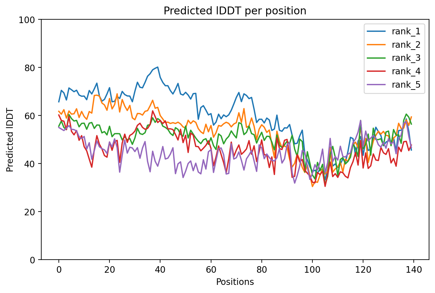
Average Confidence: 59.5%

01/3D Structure

📱 For the best experience, view 3D structures on a desktop computer.
? About the 3D Viewer

Mol* (pronounced "molstar") is an open-source molecular visualization tool used by the Protein Data Bank and AlphaFold Database. Learn more at molstar.org.

Controls:

  • Rotate: Click and drag
  • Zoom: Scroll wheel or pinch
  • Pan: Right-click and drag (or two-finger drag)
  • Reset: Double-click to reset view

What am I looking at?

This is a predicted 3D structure of the protein. The ribbon diagram shows the protein backbone—helices appear as coils, sheets as arrows, and loops as simple lines. The shape determines how the protein functions: where it binds to other molecules, how it catalyzes reactions, and how mutations might disrupt its activity.

Color legend:

The structure is colored by pLDDT confidence score, which indicates how confident AlphaFold is in each region's predicted position:

  • Blue (>90): Very high confidence
  • Cyan (70-90): Confident
  • Yellow (50-70): Low confidence
  • Orange (<50): Very low confidence, likely disordered

02/AI Analysis

TLDR

Alpha-synuclein is a protein whose clumping in brain cells causes Parkinson's disease, and the E46K mutation is a rare inherited form that makes people develop symptoms earlier and more severely. Scientists used AlphaFold2 to predict the three-dimensional structure of this mutant protein, obtaining a model with average confidence of 59.5 out of 100, indicating high uncertainty typical of disordered proteins. The low confidence reflects alpha-synuclein's naturally floppy structure, which paradoxically enables the harmful clumping behavior central to Parkinson's pathology.

Detailed Analysis

Alpha-synuclein is an intrinsically disordered protein (IDP) that lacks a stable three-dimensional structure under normal conditions but plays a central role in Parkinson's disease (PD) through its tendency to misfold and aggregate into toxic clumps called Lewy bodies [1]. The E46K mutation, where glutamic acid (negatively charged) is replaced by lysine (positively charged) at position 46, is one of five single-point mutations linked to familial Parkinson's disease and causes early-onset, aggressive forms of the condition [1]. This charge reversal fundamentally alters the protein's electrostatic properties, affecting how it interacts with itself and cellular membranes. The AlphaFold2 structural prediction yielded a model with an average confidence score (pLDDT) of 59.5, which falls well below the 70-point threshold typically used to indicate reliable structural predictions. This low confidence is expected rather than problematic, as alpha-synuclein is an intrinsically disordered protein that naturally exists as an ensemble of rapidly interconverting shapes rather than adopting a single stable fold [1]. The E46K mutation's effects are particularly sensitive to ionic strength conditions, with computational studies showing that different salt concentrations can modulate the relative aggregation propensity of mutant versus wild-type alpha-synuclein [1]. Because the predicted structure represents only one potential conformation of this dynamic protein, caution is warranted when interpreting specific structural features. The E46K mutation appears to enhance alpha-synuclein's aggregation propensity through altered electrostatic interactions, though the precise mechanisms remain under investigation [1]. Research demonstrates that alpha-synuclein pathology involves complex cellular processes including oxidative stress, mitochondrial dysfunction, and impaired autophagy-lysosome pathway activity [2][3]. The mutation likely promotes formation of aggregation-prone monomeric states that serve as precursors to the toxic oligomers and fibrils characteristic of Parkinson's disease [1]. Studies using preformed fibrils (PFFs) show that even small amounts of misfolded alpha-synuclein can seed aggregation of normal protein, creating a self-propagating cascade marked by phosphorylation at serine 129 [4]. Clinically, E46K carriers typically develop Parkinson's symptoms earlier than sporadic cases and may experience faster disease progression. The mutation exemplifies how single amino acid changes in intrinsically disordered proteins can dramatically alter disease risk and trajectory. Growing evidence suggests that Parkinson's pathology may originate in the gut, with enteric alpha-synuclein aggregation preceding brain involvement [5]. This gut-brain connection is particularly relevant for understanding how environmental factors interact with genetic mutations like E46K to trigger disease onset. The high structural uncertainty in this AlphaFold2 model reflects fundamental biology rather than technical limitations. For intrinsically disordered proteins like alpha-synuclein, understanding disease mechanisms requires studying dynamic conformational ensembles and aggregation pathways rather than static structures. Future research combining computational predictions with experimental techniques that capture protein dynamics will be essential for developing therapies targeting E46K and other familial Parkinson's mutations.

Works Cited

[1] Tammara et al. (2026). Combined Effects of Mutation and Ionic Strength on alpha-Synuclein Reveal Generic Features of Aggregation-Prone Monomeric States. ACS chemical neuroscience. [PubMed](https://pubmed.ncbi.nlm.nih.gov/42411659/) [2] Tsukiboshi et al. (2026). A PARK9 iPSC-Derived Dopaminergic Neuron Model Enables Drug Screening Targeting Autophagy-Lysosome Pathway Dysfunction in Parkinson's Disease. Journal of neurochemistry. [PubMed](https://pubmed.ncbi.nlm.nih.gov/42400323/) [3] Sayfitdinkhazhaev et al. (2026). [Parkinson's disease associated with a mutation in the glucocerebrosidase gene]. Zhurnal nevrologii i psikhiatrii imeni S.S. Korsakova. [PubMed](https://pubmed.ncbi.nlm.nih.gov/42360210/) [4] Han et al. (2026). An automated workflow for quantifying the formation of synuclein aggregates in human dopaminergic neurons. Methods (San Diego, Calif.). [PubMed](https://pubmed.ncbi.nlm.nih.gov/42297199/) [5] Giachino et al. (2026). Peripheral Immune Challenge Drives Enteric alpha-Synuclein and Tau Pathology in LRRK2 G2019S Mice. Aging and disease. [PubMed](https://pubmed.ncbi.nlm.nih.gov/42295088/)

Similar Research

**Protein quality control systems in neurodegeneration - culprits, mitigators, and solutions?** Ciechanover et al. (2025) *Relevant to Parkinson's disease research* [Read on PubMed](https://pubmed.ncbi.nlm.nih.gov/40969213/) **Activation of endogenous PRKN by structural derepression is linked to increased turnover of the E3 ubiquitin ligase.** Fiesel et al. (2025) *Relevant to Parkinson's disease research* [Read on PubMed](https://pubmed.ncbi.nlm.nih.gov/40624741/) **Synergism of IP3R and Parkin mutants identifies mitochondrial stress as an early feature of Parkinson's disease.** Dileep et al. (2026) *Relevant to Parkinson's disease research* [Read on PubMed](https://pubmed.ncbi.nlm.nih.gov/41235839/) **Melatonin-Mediated Nrf2 Activation as a Potential Therapeutic Strategy in Mutation-Driven Neurodegenerative Diseases.** Inigo-Catalina et al. (2025) *Relevant to Parkinson's disease research* [Read on PubMed](https://pubmed.ncbi.nlm.nih.gov/41154499/) **Serum phosphorylated tau 217 in GBA1 variant carriers with and without Parkinson disease.** Menozzi et al. (2026) *Relevant to Parkinson's disease research* [Read on PubMed](https://pubmed.ncbi.nlm.nih.gov/41569009/)

03/Research Data

ClinVar Classification

Not found in ClinVar

Population Frequency

No population data available

Disease Associations

801 total
Hereditary late-onset Parkinson disease
0.79
literature: 0.01 genetic association: 0.86 genetic literature: 0.88
Young adult-onset Parkinsonism
0.77
literature: 0.08 genetic association: 0.88 genetic literature: 0.88
Lewy body dementia
0.74
literature: 0.95 animal model: 0.43 genetic association: 0.82 genetic literature: 0.78
Parkinson disease
0.71
rna expression: 0.04 genetic literature: 0.78 clinical: 0.49 literature: 1.00 genetic association: 0.86
AL amyloidosis
0.46
affected pathway: 0.76

Showing 5 of 801 associations

AI Research Brief

Research brief will be generated when agent findings are available.

04/AlphaFold Metrics
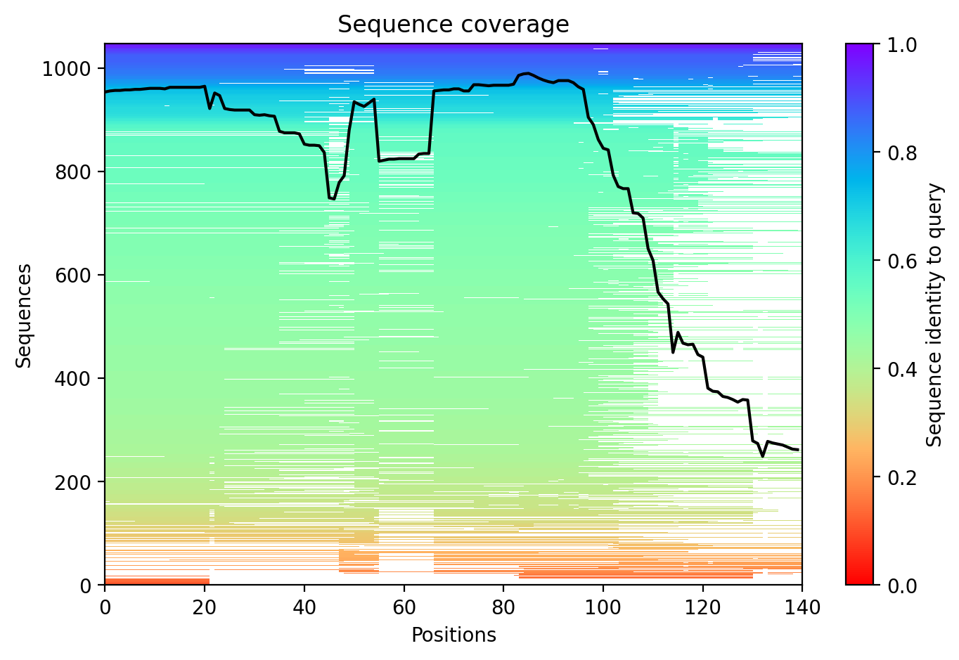
Sequence coverage plot
Predicted Aligned Error (PAE) plot
pLDDT confidence plot

05/Domain Annotations

Structural Domains & Regions

residues 20–30 Repeat — 1
residues 31–41 Repeat — 2
residues 42–56 Repeat — 3; approximate
residues 57–67 Repeat — 4
residues 20–67 Region — 4 X 11 AA tandem repeats of [EGS]-K-T-K-[EQ]-[GQ]-V-X(4)
residues 100–140 Region — Disordered
residues 111–140 Region — Interaction with SERF1A
residues 112–140 Compositional bias — Acidic residues

Functional Sites

residue 2 Binding site
residue 50 Binding site

Binding Partners

SNCAIP (22 experiments)
MAPT (12 experiments)
APOE (11 experiments)
SOD1 (9 experiments)
YWHAH (9 experiments)
PRKN (8 experiments)
TPPP (8 experiments)
HSPA1B (7 experiments)
ABL1 (6 experiments)
APP (6 experiments)

Gene Ontology

actin cytoskeleton GO:0015629 axon GO:0030424 axon terminus GO:0043679 cell cortex GO:0005938 cytoplasm GO:0005737 cytosol GO:0005829 extracellular region GO:0005576 extracellular space GO:0005615 growth cone GO:0030426 inclusion body GO:0016234 Lewy body GO:0097413 lysosome GO:0005764 membrane GO:0016020 mitochondrial inner membrane GO:0005743 mitochondrial matrix GO:0005759 +100 more

06/Structural Caption

Alpha-synuclein E46K variant shows predominantly disordered structure with modest N-terminal repeat stability; mutation disrupts electrostatic patterning in the membrane-binding domain.

Average pLDDT of 59.5 with only 19% high-confidence residues indicates a predominantly disordered protein. The N-terminal tandem repeat region (residues 20-67) shows modest stability while the C-terminal domain (residues 100-140) is highly destabilized.

The four tandem repeats (residues 20-67) correspond to the highest confidence region, consistent with lipid-binding amphipathic helices. The annotated disordered C-terminal region (residues 100-140) containing acidic residues and SERF1A interaction site shows uniformly low confidence, confirming intrinsic disorder.

The E46K mutation replaces a negatively charged glutamate with positively charged lysine within tandem repeat 3, disrupting the electrostatic balance critical for membrane binding and potentially enhancing pathological aggregation propensity characteristic of familial Parkinson's disease.

07/Peptide Therapeutics

Aggregation Analysis

Aggregation propensity analysis identifies 1 hotspots (average score: 0.00) using Pawar+KyteDoolittle+charge algorithm.

Residues 15–19 (0.51)

08/Known Inhibitors

Known Binders from ChEMBL

CHEMBL3593932 Ki: 2.1 nM (pChEMBL 8.68)

CHEMBL3593932

CHEMBL3593930 Ki: 3.5 nM (pChEMBL 8.46)

CHEMBL3593930

CHEMBL3593928 Ki: 3.8 nM (pChEMBL 8.42)

CHEMBL3593928

CHEMBL3593911 Ki: 4.2 nM (pChEMBL 8.38)

CHEMBL3593911

CHEMBL3593934 Kd: 8.9 nM (pChEMBL 8.05)

CHEMBL3593934

CHEMBL3593904 Ki: 11.5 nM (pChEMBL 7.94)

CHEMBL3593904

CHEMBL3593926 Ki: 12.9 nM (pChEMBL 7.89)

CHEMBL3593926

CHEMBL3593922 Ki: 14.6 nM (pChEMBL 7.84)

CHEMBL3593922

CHEMBL3593924 Ki: 25.0 nM (pChEMBL 7.6)

CHEMBL3593924

CHEMBL3593915 Ki: 29.8 nM (pChEMBL 7.53)

CHEMBL3593915

09/Candidate Peptides

De Novo Peptide Design Pipeline

Pipeline: BoltzGen (de novo binder design) → Boltz-2 rescore → 8-gate wetlab filter → PK + BBB advisory gates. Target site selected from UniProt curated annotations, P2Rank pocket prediction, and aggregation propensity (in that priority order). Advisory gates annotate each candidate with estimated serum half-life, renal/immunogenicity risk, and (for CNS targets) a recommended blood-brain-barrier shuttle conjugation — without silently dropping designs.

Loading candidate statistics...

Sequences are withheld pending IP review. Full candidate data (sequences, scores, CIF files) is available to authorized reviewers via the /api/private/candidates/{fold_id} endpoint with X-Private-Key.

Legacy candidates (charge-complementary)

Target Region

Residues 15–19 (0.51 aggregation score)

Candidate ID

CP-ALPHA-001 (7 residues · computational design)
âš  Drug-likeness concerns Stability: low | Toxicity: low
t½ ≈ 5 min renal high ⚙ mods suggested 🧠 Glutathione conjugate 👃 intranasal option

10/Agent Findings

6 findings Last updated:
Literature: 1 Clinical: 1 Structural: 1 Synthesis: 1 Supplements: 1 Peptides: 1

Literature Agent (1)

Literature Agent

None of the provided papers directly investigated the ALPHA-SYNUCLEIN E46K variant. While several papers examined other familial SNCA mutations (A30P, A53T) and general alpha-synuclein pathology relevant to Parkinson's disease, the specific E46K variant was not mentioned in any abstract. The papers provide useful context about alpha-synuclein biology and PD mechanisms but lack direct relevance to the E46K variant specifically.

Clinical Agent (1)

Clinical Agent

No summary available

Structural Agent (1)

Structural Agent

AlphaFold structure update: Baseline check: 3 structure(s) found

Supplements Agent (1)

Supplements Agent

Found 28 clinical trials for ALPHA-SYNUCLEIN E46K (20 recruiting). Also found 13 relevant preprints.

Synthesis Agent (1)

Synthesis Agent

Synthesis of 5 findings (clinical, literature, peptides, structural, supplements): The E46K variant of alpha-synuclein associated with Parkinson's disease presents a developing therap...

Peptide Agent (1)

Peptide Agent

ALPHA-SYNUCLEIN E46K: 10 known binders (top: 2.1 nM); 1 candidate peptides designed