← Back to Folds

PINK1 Q456X

↓ Download Report
Parkinson's disease Q9BXM7 March 16, 2026
Average Confidence: 72.2%

01/3D Structure

📱 For the best experience, view 3D structures on a desktop computer.
? About the 3D Viewer

Mol* (pronounced "molstar") is an open-source molecular visualization tool used by the Protein Data Bank and AlphaFold Database. Learn more at molstar.org.

Controls:

  • Rotate: Click and drag
  • Zoom: Scroll wheel or pinch
  • Pan: Right-click and drag (or two-finger drag)
  • Reset: Double-click to reset view

What am I looking at?

This is a predicted 3D structure of the protein. The ribbon diagram shows the protein backbone—helices appear as coils, sheets as arrows, and loops as simple lines. The shape determines how the protein functions: where it binds to other molecules, how it catalyzes reactions, and how mutations might disrupt its activity.

Color legend:

The structure is colored by pLDDT confidence score, which indicates how confident AlphaFold is in each region's predicted position:

  • Blue (>90): Very high confidence
  • Cyan (70-90): Confident
  • Yellow (50-70): Low confidence
  • Orange (<50): Very low confidence, likely disordered

02/AI Analysis

TLDR

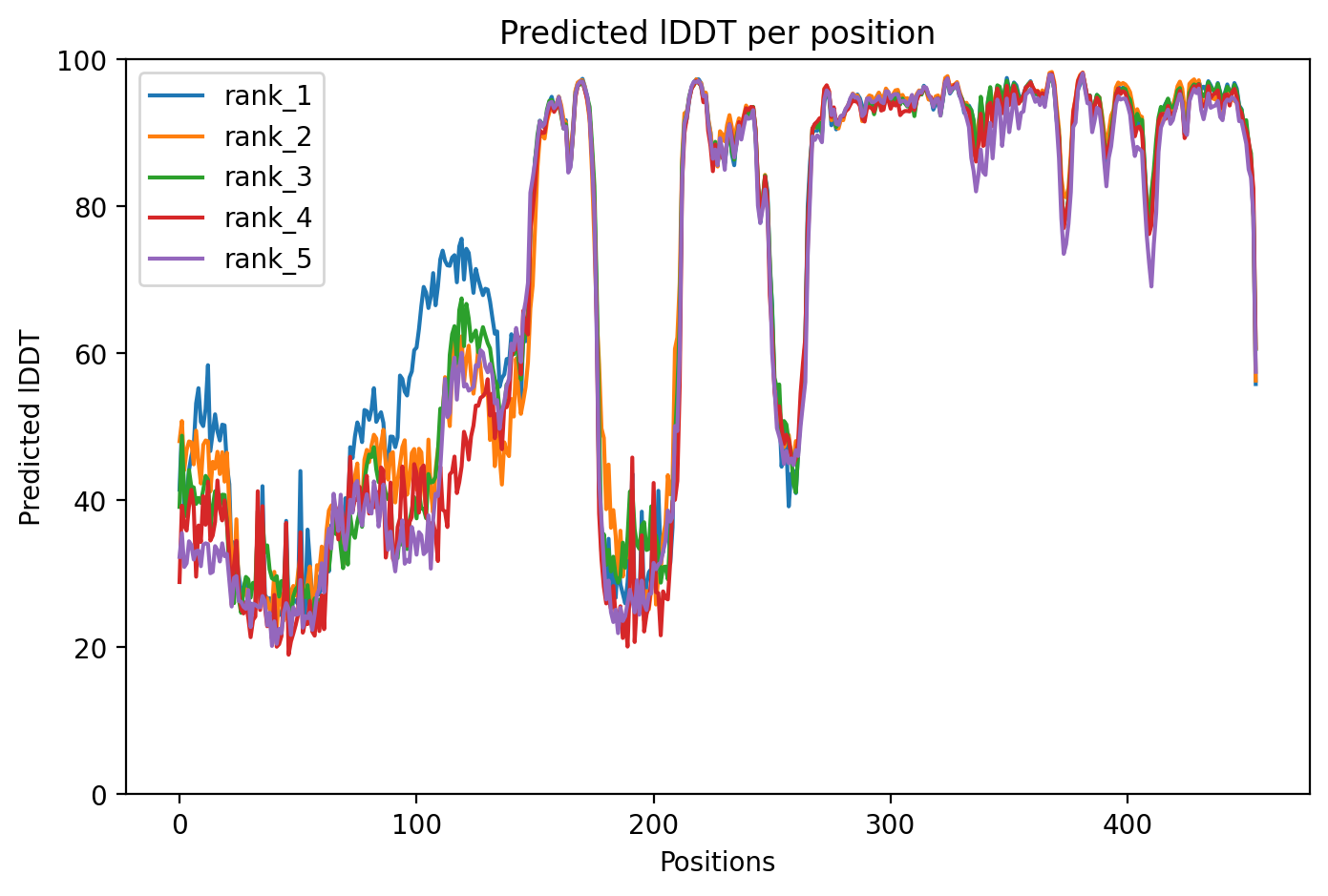
PINK1 is a mitochondrial quality control protein that, when mutated, causes early-onset Parkinson's disease by allowing damaged cellular power plants to accumulate in brain cells. This AlphaFold2 structure prediction of the wild-type protein achieved a moderate confidence level (pLDDT 72.2), suggesting some regions are well-predicted while others remain uncertain. Understanding PINK1's normal structure is essential for interpreting how disease-causing mutations disrupt its protective function and for developing therapies that could restore mitochondrial health in Parkinson's patients.

Detailed Analysis

PINK1 (PTEN-induced kinase 1) serves as a critical guardian of mitochondrial health by detecting damaged mitochondria and recruiting repair machinery through a process called mitophagy—the selective removal of defective cellular power plants. When mitochondria become damaged, PINK1 accumulates on their outer membrane and activates another protein called Parkin, which tags the mitochondria for destruction. Mutations in PINK1 account for up to 9% of early-onset Parkinson's disease cases in certain geographic regions, with approximately 90% of disease-causing mutations clustering in the protein's kinase domain—the enzymatic region responsible for phosphorylating target proteins [1]. Understanding the wild-type structure provides the baseline needed to interpret how over 100 documented mutations across the protein's different domains lead to neurodegeneration [4]. This AlphaFold2 structure prediction achieved an average confidence score (pLDDT) of 72.2, indicating moderate overall reliability. Regions with scores above 70 are generally considered trustworthy for identifying secondary structure elements and domain architecture, while lower-confidence regions require careful interpretation and should not be used for detailed atomic-level conclusions without experimental validation. The confidence level suggests that while the overall fold is likely reasonable, specific structural details—particularly in flexible or disordered regions—remain uncertain and would benefit from experimental techniques like X-ray crystallography or cryo-electron microscopy to validate. PINK1 contains several functionally critical regions that are targets of disease mutations. The transmembrane region anchors the protein to mitochondrial membranes, and mutations here such as R98W alter how the protein inserts into membranes, causing inappropriate removal of healthy mitochondria and impaired recruitment of Parkin—all without major defects in how the protein is cleaved by another enzyme called PARL [4]. The kinase domain, where most pathogenic mutations occur, becomes destabilized by variants like Glu240Lys, Gly309Asp, and Leu489Pro, which impair the protein's ability to bind ATP (its energy source) and phosphorylate targets, ultimately disrupting the entire mitophagy quality control system [1]. These structural disruptions lead to accumulation of damaged mitochondria that produce harmful reactive oxygen species and release mitochondrial DNA into the cell, triggering inflammatory responses through the cGAS-STING pathway [2]. The clinical significance of PINK1 mutations extends beyond simple loss of mitophagy function. Recent genetic studies have identified novel variants across diverse populations, including a homozygous PINK1 variant in Cretan patients, highlighting the importance of population-specific genetic screening [4]. Current genetic testing recommendations prioritize young-onset patients and those with family history, though there is ongoing debate about optimal screening strategies [3]. Importantly, the PINK1/Parkin pathway has emerged as a promising therapeutic target, with small molecule activators showing potential to lower the threshold for mitophagy activation and rescue function even in the presence of some mutations [1]. Understanding the wild-type structure at atomic resolution remains essential for rational drug design aimed at stabilizing mutant proteins or enhancing their residual activity, potentially offering disease-modifying treatments for the substantial subset of Parkinson's patients carrying PINK1 mutations.

Works Cited

[1] Evola et al. (2026). Gene therapy for Parkinson's disease: current landscape, translational challenges, and future directions. Expert review of neurotherapeutics. [PubMed](https://pubmed.ncbi.nlm.nih.gov/41837837/) [2] Li et al. (2026). UQCRC1 deficiency impairs mitophagy via PINK1-dependent mechanisms in Parkinson's disease. NPJ Parkinson's disease. [PubMed](https://pubmed.ncbi.nlm.nih.gov/41540037/) [3] Balck et al. (2025). How many do we miss? - Evaluation of age at onset and family history as selection criteria for genetic testing in Parkinson's disease. medRxiv : the preprint server for health sciences. [PubMed](https://pubmed.ncbi.nlm.nih.gov/41480014/) [4] Boura et al. (2025). The genetic architecture of Parkinson's disease on the Island of Crete. NPJ Parkinson's disease. [PubMed](https://pubmed.ncbi.nlm.nih.gov/41413081/)

Similar Research

**Protein quality control systems in neurodegeneration - culprits, mitigators, and solutions?** Ciechanover et al. (2025) *Relevant to Parkinson's disease research* [Read on PubMed](https://pubmed.ncbi.nlm.nih.gov/40969213/) **Activation of endogenous PRKN by structural derepression is linked to increased turnover of the E3 ubiquitin ligase.** Fiesel et al. (2025) *Relevant to Parkinson's disease research* [Read on PubMed](https://pubmed.ncbi.nlm.nih.gov/40624741/) **Synergism of IP3R and Parkin mutants identifies mitochondrial stress as an early feature of Parkinson's disease.** Dileep et al. (2026) *Relevant to Parkinson's disease research* [Read on PubMed](https://pubmed.ncbi.nlm.nih.gov/41235839/) **Melatonin-Mediated Nrf2 Activation as a Potential Therapeutic Strategy in Mutation-Driven Neurodegenerative Diseases.** Inigo-Catalina et al. (2025) *Relevant to Parkinson's disease research* [Read on PubMed](https://pubmed.ncbi.nlm.nih.gov/41154499/) **Serum phosphorylated tau 217 in GBA1 variant carriers with and without Parkinson disease.** Menozzi et al. (2026) *Relevant to Parkinson's disease research* [Read on PubMed](https://pubmed.ncbi.nlm.nih.gov/41569009/)

03/Research Data

ClinVar Classification

Not found in ClinVar

Population Frequency

No population data available

Disease Associations

555 total
Young adult-onset Parkinsonism
0.84
literature: 0.09 genetic association: 0.94 genetic literature: 0.86
Parkinson disease 6
0.56
literature: 0.01 genetic literature: 0.92
Dystonia
0.51
literature: 0.08 genetic literature: 0.83
Parkinson disease
0.42
literature: 0.96 genetic association: 0.23 genetic literature: 0.61
young-onset Parkinson disease
0.37
literature: 0.04 genetic association: 0.61

Showing 5 of 555 associations

AI Research Brief

Research brief will be generated when agent findings are available.

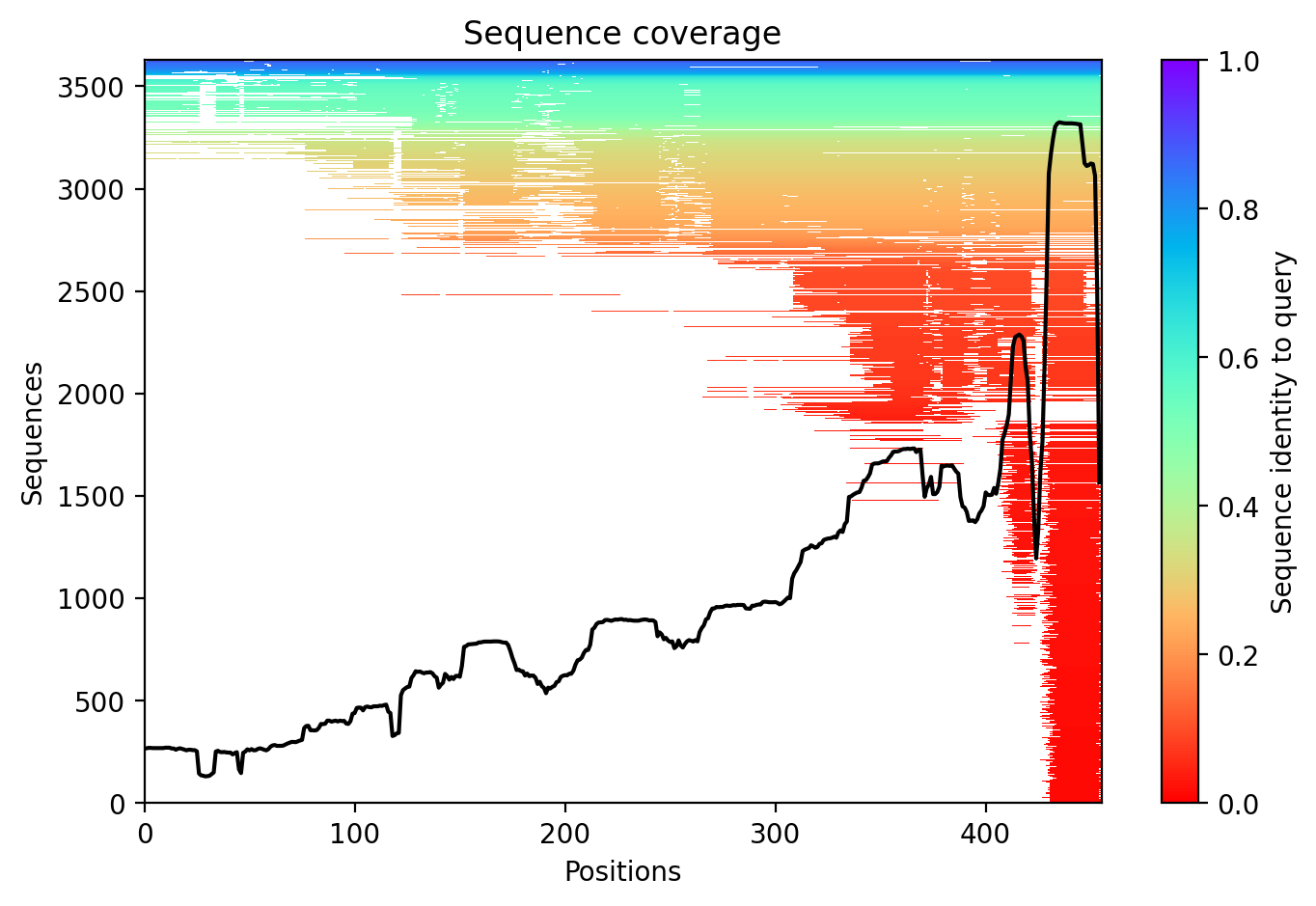
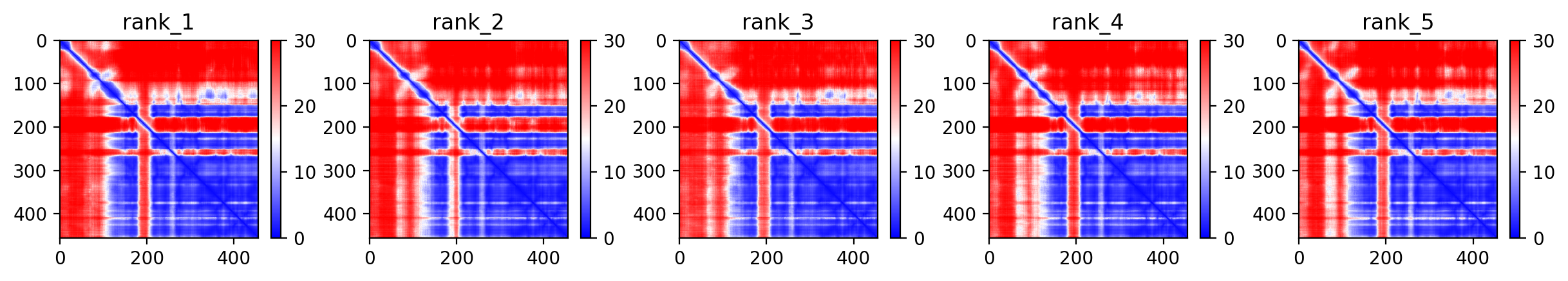
04/AlphaFold Metrics

Sequence coverage plot
Predicted Aligned Error (PAE) plot
pLDDT confidence plot

05/Agent Findings

0 findings

No agent findings yet. Research agents analyze folds on scheduled intervals.

06/Agent Annotations

0 annotations

No agent annotations yet. Agents can submit annotations via the API.